
Facets
Browse across eight MeSH (opens in new tab) facets — era, geography, science, specialty, technology, history, culture, and reference. Select one tag per group; counts update across the others.
Clear filtersFacet filters
Geography
Specialties & Disease
- Anatomy & Pathology 290
- Cardiology & Blood 3
- Neurology & Psychiatry 19
- Obstetrics & Reproductive 3
- Infectious Disease (General) 0
- Surgery & Anesthesia 14
- Public Health 6
- Immunology & Dermatology 11
- General Clinical Medicine 4
- Military Medicine 0
- Psychology 1
- Alternative & Fringe Medicine 0
- Pediatrics 2
- Ophthalmology & Vision 4
- ENT & Hearing 5
- Urology & Nephrology 12
- Gastroenterology & Hepatology 20
- Pulmonary & Respiratory 1
- Rheumatology, Rehab & Pain 0
- Internal, Emergency & Geriatric 0
- Veterinary Medicine 1
- Epidemiology & Demography 0
- Physiology & Embryology 29
- Dentistry 4
- Plagues & Epidemics 0
- Microbiology & Virology 3
Social & Historical Studies
Institutions & Culture
Reference & Scholarly Works
Drugs & Technology
19 entries match Modern [K01.400.504] · Neurology & Psychiatry [C10 / F04]
1992 CE
#7354
Brain maps: Structure of the rat brain.
The first computer graphics atlas of the brain of any species, with the illustration files also available separately (1993). The work included a complete and systematic, hierarchically organized set of annotated nomen…
1836 CE–1842 CE
#13300
A series of anatomical plates. 5 vols.: The muscles of the human body. The vessels of the human body. The nerves of the human body. The viscera of the human body. The bones and ligaments of the human body.
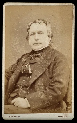
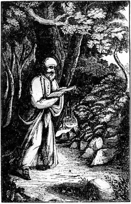
The most ambitious 19th century English anatomy illustrated by lithography. Some copies were issued with hand-colored plates. The five volumes, containing a total of 201 plates, describe the muscles, blood vessels, ne…
1943 CE
#7347
An atlas of the basal ganglia, brain stem and spinal cord, Based on myelin-stained material.
"Classic atlas of the human brain (excluding the cerebral and cerebellar cortex). Each structure has a blurb with varying amounts of useful historical and factual information. There is also a very useful bibliography …
1611 CE
#6964
Anatomicae institutiones corporis humani.
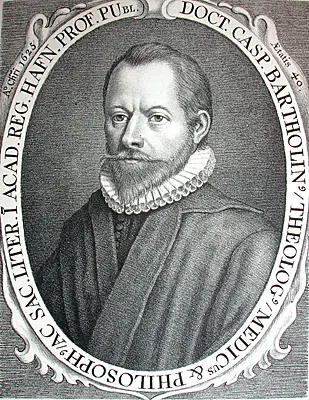
The elder Caspar Bartholin was the first to describe the workings of the olfactory nerve, and introduced the terms nervus olfactorius and nervus vagus. This was a standard textbook for many years, undergoing numerous …
2016 CE
#11138
Anatomy: An encyclopedic reference to the language of anatomy and neuroanatomy. It provides the fascinating origin of terms and biographies of anatomists/physicians who originated them.
1861 CE–1867 CE
#14159
Atlas des peripherischen Nervensystems des menschlichen Körpers / Atlas du système nerveux périphérique du corps humain. Mit einem Vorwort von Prof. Dr. Th. W. L. Bischoff, nach der Natur photographirt von Joseph Alb. 10 parts.
Issued in 10 parts in folio (510 x 360 mm). Includes 46 large-format original photographs mounted on sheets of cardboard.
1893 CE
#431
Atlas of head sections. Fifty-three engraved copperplates of frozen sections of the head, and fifty-three key plates with descriptive texts.
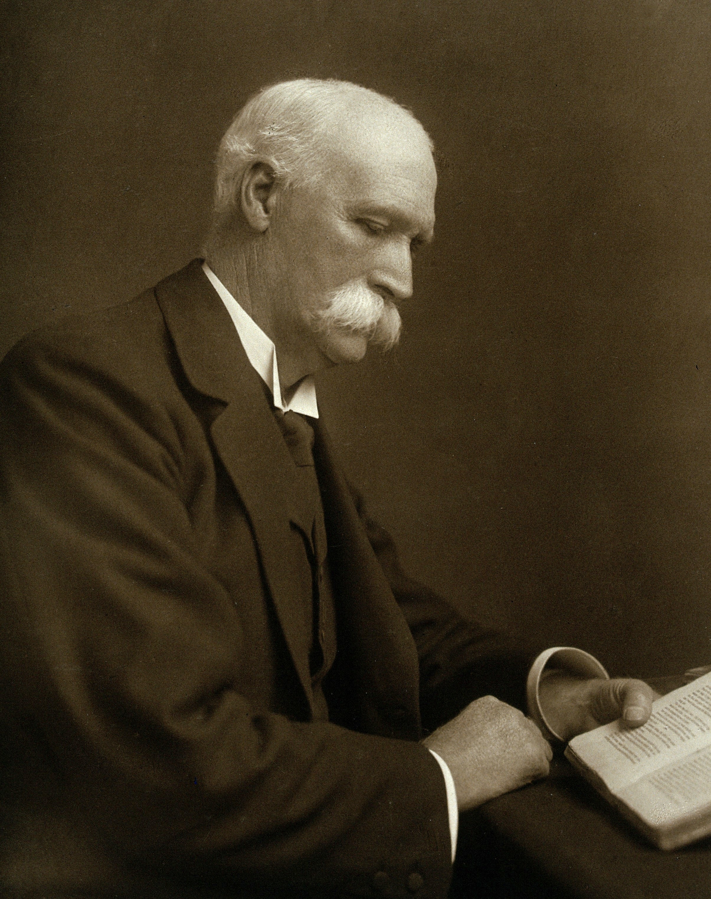
Intended to supplement and illustrate Macewen’s neurosurgical textbook published the same year (No. 4872). Includes coronal, sagittal and horizontal sections with commentary on each.
2012 CE
#9418
Atlas of human brain connections.
The authors combined the science of diffusion tensor imaging with the art of tractography: Spectacular color images.
1971 CE
#7353
Atlas of the mouse brain and spinal cord.
"This is an exceptionally systematic, beautifully produced atlas of the mouse central nervous system in the three standard planes, with alternating cell-stained (Nissl method with cresyl violet) sections and myelin-st…
1732 CE
#1314
Exposition anatomique de la structure du corps humain.
![WINSLOW, Jacques-Bénigne [Jacob Benignus Winsløw] (1669 – 1760)](/media/portraits/708)
The foramen between the greater and lesser sacs of the peritoneum (described on pages 352-65), is named after Winslow. His Exposition is distinguished as being the first book on descriptive anatomy to discard physiolo…
1684 CE
#1379
Neurographia universalis.
Vieussens, professor at Montpellier, was the first to describe the centrum ovale correctly. The publication of the above work threw new light on the subject of the configuration and structure of the brain, spinal cord…
1840 CE
#1263
Nuovi organi scoperti nel corpo humano.
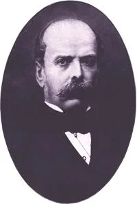
“Pacini’s corpuscles”, end organs of sensory nerves, earlier described by Vater in 1717.
1714 CE
#1312
Tabulae anatomicae.
A romantic history attaches to this fine collection of plates, drawn by Eustachius himself and completed in 1552. They remained unprinted and forgotten in the Vatican Library until discovered in the early 18th century…
1998 CE
#7350
The central nervous system of vertebrates. 3 vols.
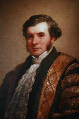
A massive contribution to comparative vertebrate neuroanatomy, the life-work of the authors. Includes a comprehensive account of the structural organisation of all vertebrate groups, ranging from amphioxus and lamprey…
1864 CE
#7331
The gray substance of the medulla oblongata and trapezium.
The first American medical book illustrated with photomechanically reproduced plates. Oliver Wendell Holmes praised the book for its remarkable photomicrographs, which may be the first published of brain cross-section…
1954 CE
#7351
The human brain in sagittal section.
A superb atlas is based on sagittal sections. This was an innovative approach for the time as almost all previous illustration of the adult human brain was typically based on frontal or horizontal sections: “in …
1961 CE
#7352
The human cerebellum. An atlas of gross topography in serial sections.
1801 CE–1803 CE
#1315
Traité d’anatomie descriptive. 5 vols.
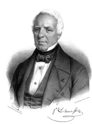
Bichat was the creator of descriptive anatomy. He introduced the terms “animal” and “vegetative” system. This was his last work, unfinished at his death. Vol. 4 was prepared by Bichat's student…
1786 CE
#401.2
Traité d’anatomie et de physiologie avec des planches coloriées répresentant au naturel les divers organes de l’homme et des animaux. Tome premier [all published].
The most accurate neuroanatomical work produced before the advent of microscopic staining techniques. Vicq d’Azyr identified accurately for the first time many of the cerebral convolutions, along with various in…