
Facets
Browse across eight MeSH (opens in new tab) facets — era, geography, science, specialty, technology, history, culture, and reference. Select one tag per group; counts update across the others.
Clear filtersFacet filters
Geography
Specialties & Disease
- Anatomy & Pathology 290
- Cardiology & Blood 3
- Neurology & Psychiatry 19
- Obstetrics & Reproductive 3
- Infectious Disease (General) 0
- Surgery & Anesthesia 14
- Public Health 6
- Immunology & Dermatology 11
- General Clinical Medicine 4
- Military Medicine 0
- Psychology 1
- Alternative & Fringe Medicine 0
- Pediatrics 2
- Ophthalmology & Vision 4
- ENT & Hearing 5
- Urology & Nephrology 12
- Gastroenterology & Hepatology 20
- Pulmonary & Respiratory 1
- Rheumatology, Rehab & Pain 0
- Internal, Emergency & Geriatric 0
- Veterinary Medicine 1
- Epidemiology & Demography 0
- Physiology & Embryology 29
- Dentistry 4
- Plagues & Epidemics 0
- Microbiology & Virology 3
Social & Historical Studies
Institutions & Culture
Reference & Scholarly Works
Drugs & Technology
290 entries match Modern [K01.400.504] · Anatomy & Pathology [G02.149 / C23]
1685 CE
#1101
De ductu salivali novo, saliva, ductibus oculorum aquosis, et humore oculi aqueo.
Nuck’s name has been attached to the glands and duct described by him.
1745 CE
#978
De fabrica et actione villorum intestinorum tenuium hominis.
“Lieberkühn’s glands” or “crypts” described. They were discovered by Malpighi in 1688.
1687 CE
#975
De glandulis in intestino duodeno hominis detectis.
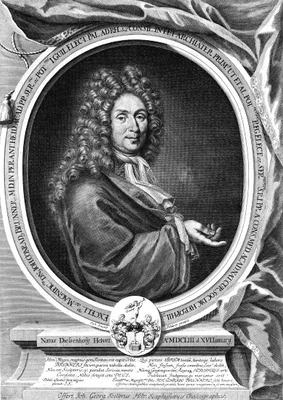
“Brunner’s glands”, earlier described by Wepfer (No. 974.1).
1652 CE
#1096
De lacteis thoracicis in homine brutisque.
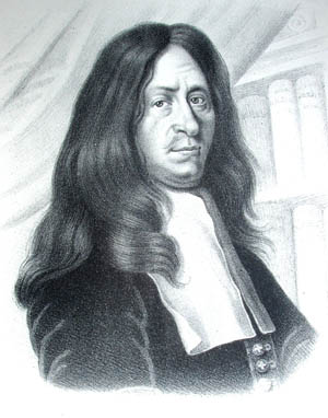
Contains Bartholin’s discovery of the thoracic duct. English translation, 1653.
1627 CE
#1094
De lactibus sive lacteis venis.
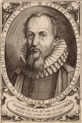
Records the discovery of the lacteal vessels. While performing vivisection on a dog that had recently fed, Aselli noticed a network of vessels in the mesentery and along the peritoneal surface of the intestine. The ve…
1672 CE
#1209
De mulierum organis generationi inservientibus.
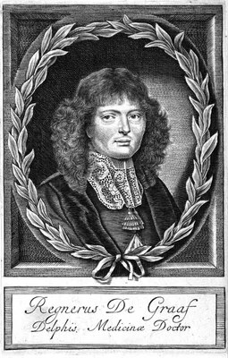
De Graaf demonstrated ovulation anatomically, pathologically and experimentally. In the above work he included the first account of the “Graafian follicle”. Translation of Chapter XII, dealing with the ova…
1661 CE
#760
De pulmonibus observations anatomicae.
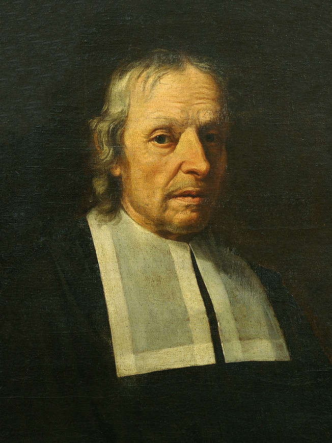
Discovery of the capillary circulation. Malpighi demonstrated that the pulmonary tissues are vesicular in nature and showed that the trachea ends in bronchial filaments. His De pulmonibus includes his demonstration of…
1707 CE
#1216
De sterilitate mulierum.
The Nabothian cysts and glands of the cervix uteri first described (sect. xv).
1603 CE
#757
De venarum ostiolis.
Fabricius, teacher of Harvey at Padua, discovered the venous valves, and illustrated them in life-size copperplates in this monograph. He failed to recognize their true function, however, considering their function si…
1668 CE
#1210
De virorum organis generationi inservientibus, de clysteribus et de usu siphonis in anatomia.
Exact and detailed account of the male reproductive system. This work and No. 1209 were translated into English and published as Suppl. 17 to J. Reprod. Fertil, 1972. Facsimile of originals, Nieuwkoop, De Graaf, 1965.
1858 CE
#420
Des moyens chirurgicaux de favoriser la reproduction des os après les résections.
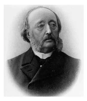
“Ollier’s layer”, the osteogenetic layer of the periosteum.
1755 CE
#1484
Descriptio anatomica oculi humani.
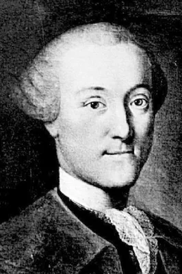
The first complete study of the anatomy of the eye, including the first description of the “zonule of Zinn” and the “annulus of Zinn”.
1826 CE
#7511
Descriptio musei anatomici.
Bleuland's catalogue of his museum of anatomical and pathological specimens. Digital facsimile from Universiteit Utrecht at this link.
1719 CE
#1215
Description de l’urèthre de l’homme.
“Littre’s glands” described.
1893 CE–1903 CE
#10367
Descriptive catalogue of the anatomical and pathological specimens in the museum of the Royal College of Surgeons of Edinburgh. 3 Vols. Vols. 1 & 2 by Charles W. Cathcart; Vol. 3 by Theodore Shennan. Vol. 1.- The skeleton and organs of motion.
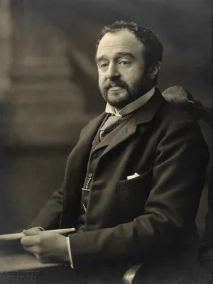
Digital facsimile of vol. 1 from the Biodiversity Heritage Library at this link.
1834 CE–1840 CE
#10380
Descriptive catalogue of the preparations in the Museum of the Royal College of Surgeons in Ireland. Vol. I. (Anatomy). Vol. 2. (Pathology).
Digital facsimile from the Internet Archive at this link.
1753 CE
#7800
Dictionaire anatomique suivi d'une bibliothèque anatomique et physiologique.
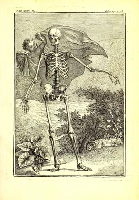
An anatomical dictionary with most of the entries cross referenced to related structures, followed by a very extensive bibliography of anatomical and physiological works. Digital facsimile from Google Books at this link.
1913 CE–1914 CE
#435
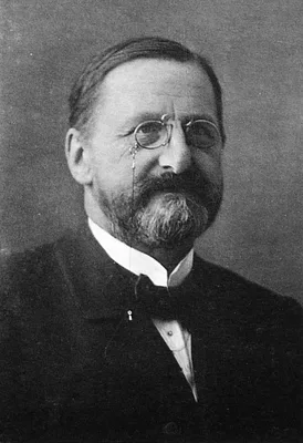
Die Anatomie des Menschen. 3 pts.
1895 CE
#432
Die anatomische Nomenclatur.
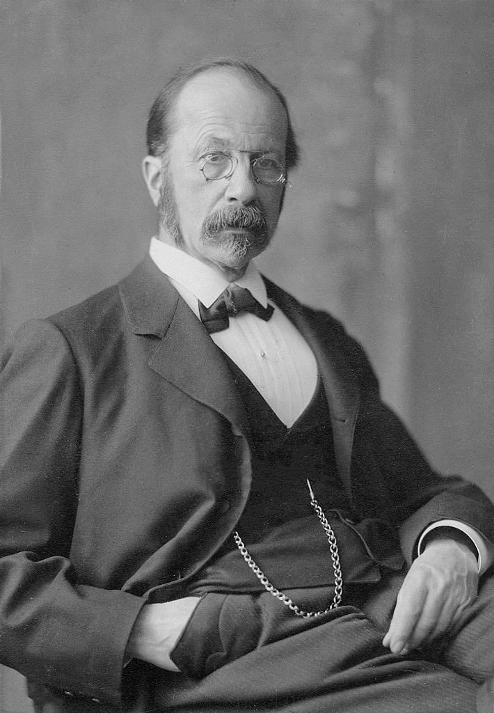
His was largely responsible for the Basle Nomina Anatomica, the first attempt to produce a standard anatomical nomenclature. English translation by L.F. Barker, 1907.
1873 CE
#425
Die Corrosions-Anatomie und ihre Ergebnisse: mit 18 chromolithographirten Tafeln.
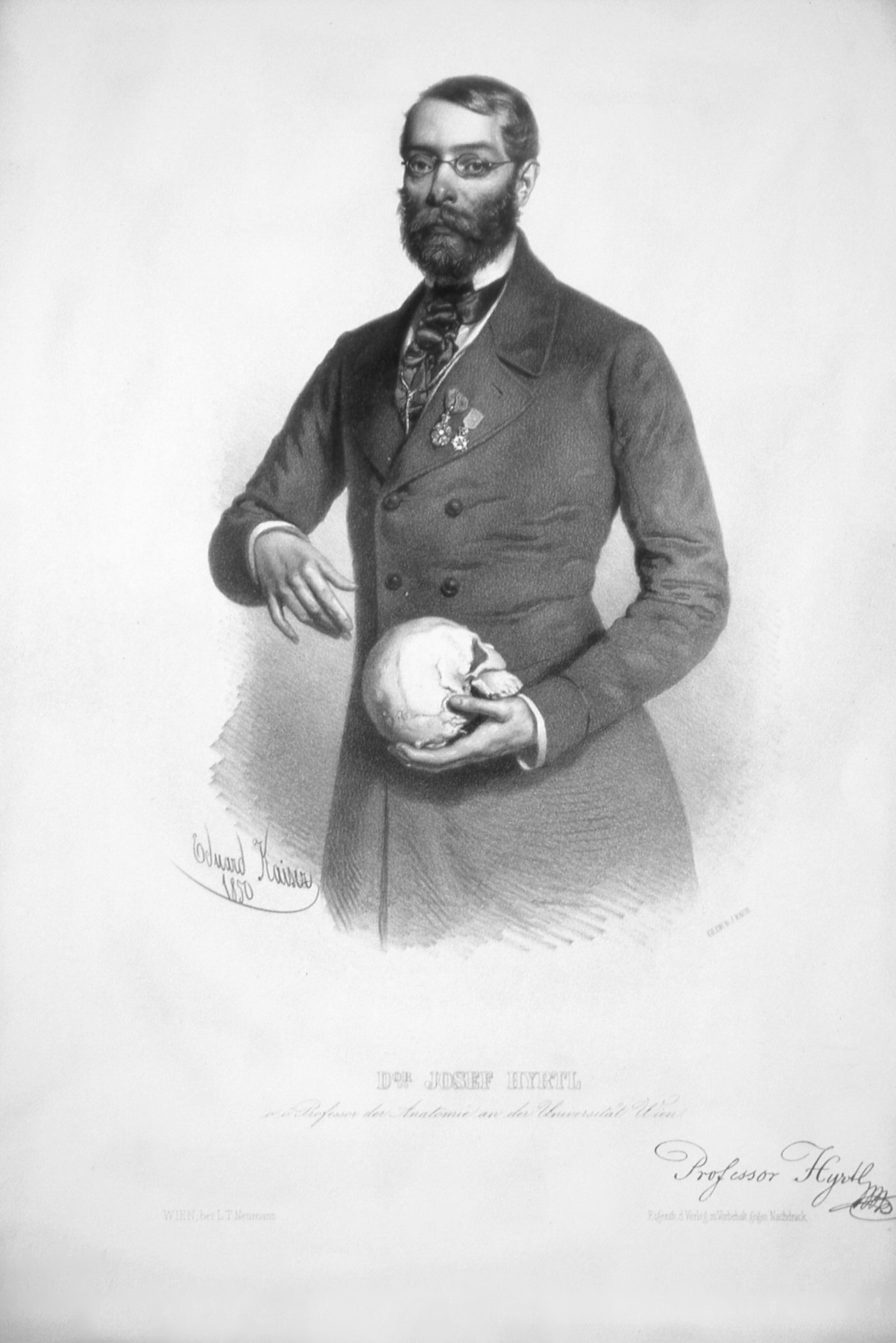
Hyrtl significantly enhanced the techniques of corrosion anatomy, a technique of preparing anatomical specimens invented by Frederik Ruysch. He built up a collection unsurpassed in Europe. In this work Hyrtl described…
1870 CE
#423
Die descriptive und topographische Anatomie des Menschen.
1872 CE
#6188
Die Lage des Uterus und Foetus am Ende der Schwangerschaft nach Durchschnitten an gefrornen Cadavern
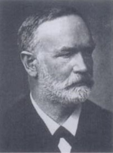
Supplement to No. 424.
1862 CE
#1108
Die Lymphgefässe und ihre Beziehung zum Bindegewebe.
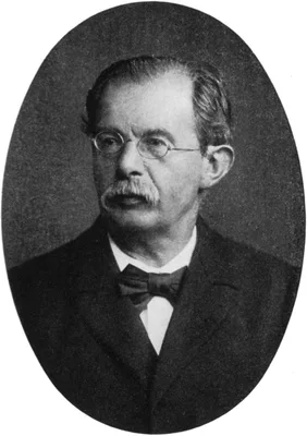
“Recklinghausen’s canals”, the lymph canaliculi.
1844 CE
#11148
Die männlichen und weiblichen Wollust-Organe des Menschen und einiger Säugetiere.
Kobelt provided the first comprehensive and accurate description of the function of the clitoris.Digital facsimile from digi.ub.uni-heidelberg at this link.
1876 CE
#2304
Die Sections-Technik im Leichenhause des Charité-Krankenhauses.
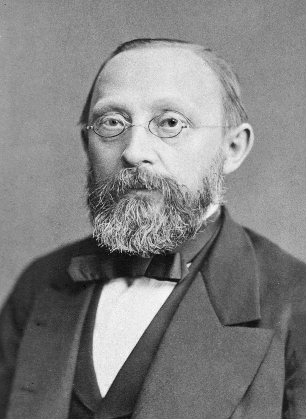
On the technique of dissection. English translation, London, 1876.
1665 CE
#1099
Dilucidatio valvularum in vasis lymphaticis et lacteis.
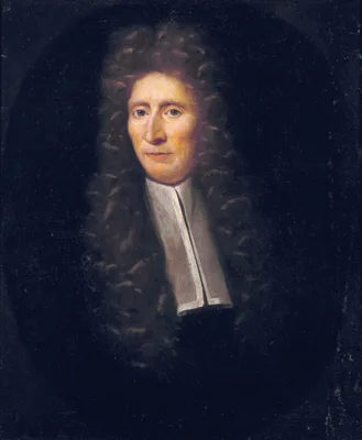
First description of the valves of the lymphatics, discovered by Ruysch. Facsimile reprint, Niewkoop, De Graaf, 1964.
1708 CE
#763
Disputatio medica inauguralis de circulo sanguinis in corde.
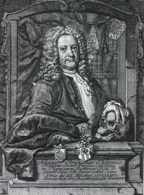
First description of the coronary valves and the venae thebesii.
1892 CE–1895 CE
#429
Dissections illustrated.
“Brodie’s ligament”, the transverse humoral ligament, described.
1720 CE
#976
Dissertatio anatomica qua novum bilis diverticulum circa orificium ductus choledochi ut et valvulosam colli vesicae felleae constructionem ad disceptandum proponit.
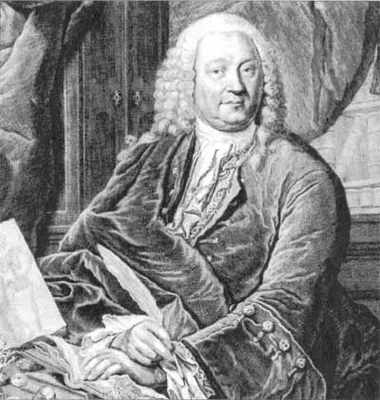
Following Vater’s classic description of the ampulla of the bile duct, it was named the “ampulla of Vater”.
1736 CE
#7507
Dissertatio de arteries et venis intestinorum hominis. Adjecta icon coloribus distincta.
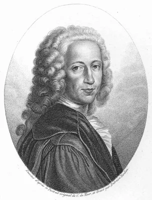
The small color mezzotint printed by the painter Jan Ladmiral included with this pamphlet on the arteries and veins of the human intestine was among the earliest applications of full color printing, and the first use …
1743 CE
#765.1
Dissertationem inauguralem de vera nervi intercostalis origine.
Taube described the carotid body and named it “ganglion minutum”, See J. Pick, J. Hist. Med., 1959, 14, 61-73.
1684 CE
#974.2
Du ductu salivati hactenus non descriptio.
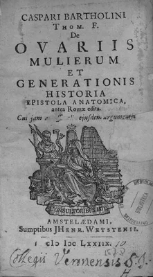
“Bartholin’s duct” and “gland”, the sublingual salivary gland and ducts.
1828 CE
#410
Elements of descriptive and practical anatomy.
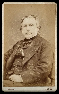
Among the most important of the English textbooks on anatomy. An eleventh edition was published in 1908-29.
1745 CE
#398
Essai d’anatomie en tableaux imprimés, qui represent au naturel tous les muscles de la face, du col, de la tête, de la langue & du larinx. d'après les parties disséquées & préparées par Monsieur Duverney....comprenant hit grandes planches.
Remarkable for its striking mezzotints printed in color. Digital facsimile from the Internet Archive at this link.
1742 CE
#396
Essais anatomiques.
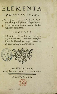
Lieutaud rectified many anatomical errors, described carefully the structure and relations of the heart and its cavities, and added to the contemporary knowledge concerning the bladder. The trigonum vesicae is named &…
1861 CE
#7608
Essays and observations on natural history, anatomy, physiology, psychology, and geology by John Hunter, F.R.S. Being his posthumous papers on those subjects, arranged and revised, with notes; to which are added the introductory lectures on the Hunterian collection of fossil remains delivered in the theatre of the Royal College of Surgeons of England, March 8th, 10th and 12th, 1855 by Richard Owen .... 2 vols.
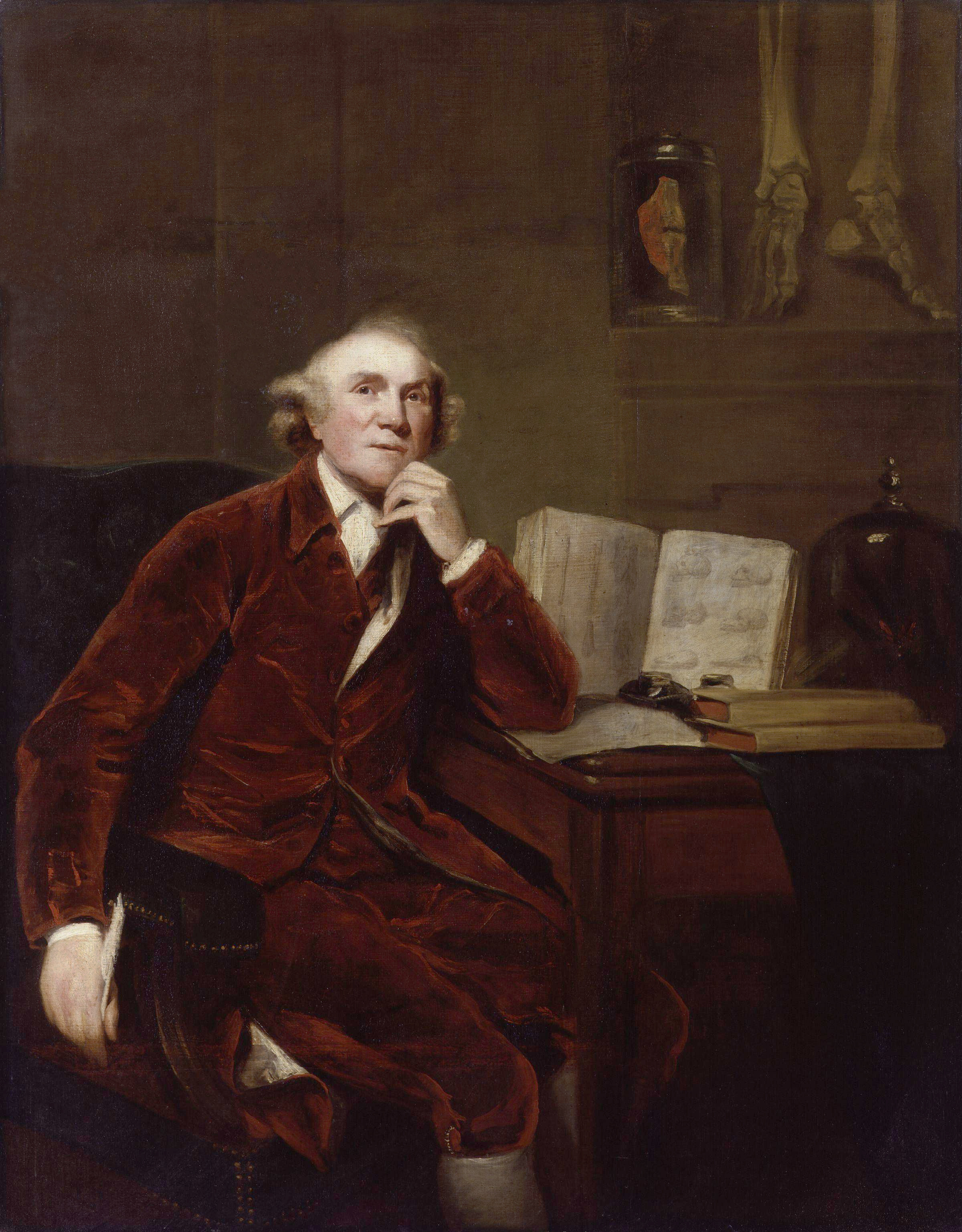
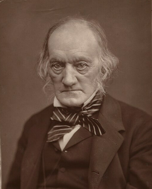
Digital facsimiles from the Hathi Trust at this link.
1677 CE
#1100
Exercitatio anatomico-medica de glandulis intestinorum, earumque usu et affectionibus.
Independently of Bartholin and Rudbeck, George Joyliffe (1621-58) observed the lymphatics. He communicated his discovery to Glisson early in 1652 and the latter included an account in the above work (Cap. xxxi). See N…
1651 CE
#1095
Experimenta nova anatomica, quibus incognitum chyli receptaculum, et ab eo per thoracem in ramos usque subclavis vasa lactea deteguntur.
Pecquet discovered the thoracic duct in dogs and its relation to the lacteals. Using a dog that was digesting, he described the thoracic duct, its entry into the subclavian veins, and the receptaculum chyli or chyle r…
1774 CE
#1102
Experimental inquiries: Part the second. Containing a description of the lymphatic system in the human subject and in other animals. Together with observations on the lymph, and the changes which it undergoes in some diseases.
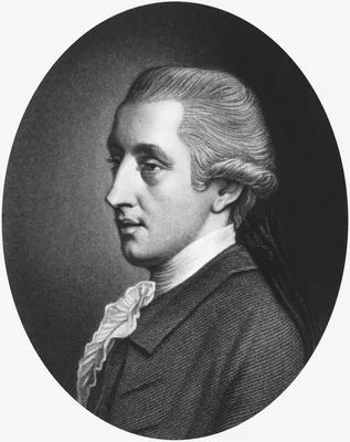
Hewson gave the first complete account of the anatomical peculiarities of the lymphatics. He divided the lymphatics into two groups – superficial and deep. He described the leucocytes as derived from the lymphat…
1732 CE
#1314
Exposition anatomique de la structure du corps humain.
![WINSLOW, Jacques-Bénigne [Jacob Benignus Winsløw] (1669 – 1760)](/media/portraits/708)
The foramen between the greater and lesser sacs of the peritoneum (described on pages 352-65), is named after Winslow. His Exposition is distinguished as being the first book on descriptive anatomy to discard physiolo…
1642 CE
#7879
Figura ductus cuiusdam cum multiplicibus suis ramulis noviter in pancreate in diversis corporibus humanis observati.
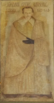
Wirsung, assistant to the celebrated German anatomist Johann Vesling, discovered the excretory duct of the pancreas named for him in 1642. To announce his discovery, Wirsung chose the extremely unusual method of publi…
2011 CE
#7005
Fragonard Museum. The écorchés. The anatomical masterworks of Honoré Fragonard by Christophe Degueurce. With an essay by Laure Cadot. Translated from the French by Philip Adds.
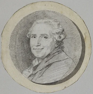
The painter and printmaker Fragonard preserved the results of his dissections via means never divulged, but which may have been based on those of Jean-Joseph Sue. His pieces were often prepared for theatrical effect r…
1881 CE
#7645
Frozen sections of a child by Thomas Dwight. Fifteen drawings from nature by H. P. Quincy.
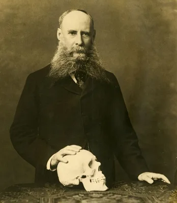
The first atlas of cross-sectional anatomy published in the United States. Digital facsimile from the Internet Archive at this link
2007 CE
#14074
Gustaf Retzius: A Biography by Thomas Lindblad. With special contributions by Gunnar Grant, Björn Afzelius, Olle Johansson & Markku Virtanen, Helge Rask-Andersen, Torstein Sjøvold. Editor: Ove Hagelin.
A finely written and superbly illustrated and produced study of Retzius's life and published works, issued in the style of Retzius's magnificent publications.
1970 CE
#14216
Hammond's wall atlas of human anatomy.
With pages measuring 731 x 536 mm., this is the largest anatomical atlas ever published with plastic overlays. The covers include metal grommets so that the book could be hung on the wall. The work was undated, but is…
1820 CE
#9696
Hamse-i Şanizade. Miratü'l Ebdan fi Teşrih-i Azaü'l-İnsan [About anatomy]; Usulü't Tabia [Book of physiology]; Miyaru'l-Etibba [Practice of medicine].
This illustrated compendium of anatomy, physiology, and internal medicine was first medical work in Turkish printed by letterpress in the Ottoman Empire. It was also one of the first medical works in Turkish to draw t…
1895 CE–1903 CE
#430
Handatlas der Anatomie des Menschen. 3 vols.
16th edition in English, 1967.
1896 CE–1934 CE
#433
Handbuch der Anatomie des Menschen. 32 parts.
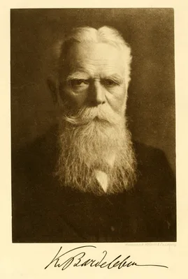
An important collective work.
1815 CE–1820 CE
#407
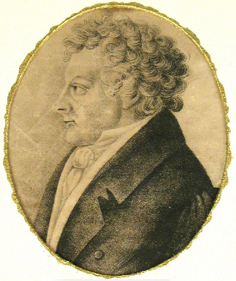
Handbuch der menschlichen Anatomie. 4 vols.
1927 CE–1943 CE
#563